CME Accredited Surgical Training Available Online

Mackool Online CME

  • Home
  • Archived Surgical Videos
    • 2016 Videos
    • 2017 Videos
    • 2018 Videos
    • 2019 Videos
    • 2020 Videos
    • 2021 Videos
    • 2022 Videos
  • Home
  • 2016 Videos
  • Archive from category "2016 Videos"
May 25, 2022

Category: 2016 Videos

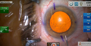
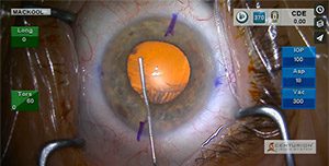
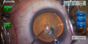
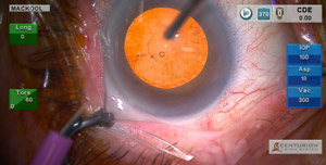
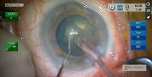
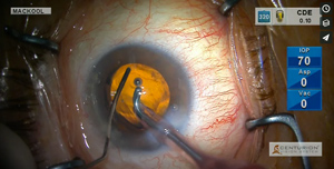
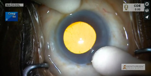
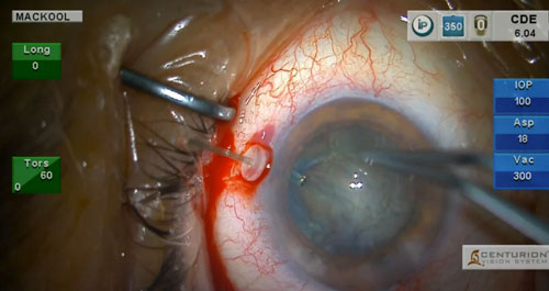
Episode 12: Advanced Capsule Retractors for Zonular Laxity

Wednesday, 14 December 2016 by Captured Light Studio

episode12thumb
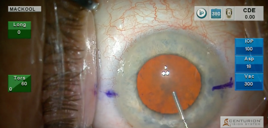
This patient has pseudoexfoliation, shallow anterior chamber, dense nucleus, lax zonule and poor pupillary dilation. I have previously demonstrated the use of capsular retractors for zonular laxity, but here we will see the latest version of these devices as  modified by Dr David Chang.  I also discuss the causes of chopping difficulties, demonstrate “verticalizing” the chopper, use of a CTR, and discuss IOL selection based on intraoperative findings.

Read more
  • Published in 2016 Videos
No Comments

Episode 11: Full Thickness (Penetrating) LRIs to Correct Astigmatism During Multifocal IOL Insertion

Monday, 14 November 2016 by Captured Light Studio

episode11_thumbnailDuring this case I demonstrate the differences between phaco aspiration vs phacoemulsification of the nucleus, as well as the use of Penetrating Limbal Relaxing Incisions to control astigmatism in a multifocal IOL patient. The PLRI nomograms may be downloaded by clicking on the “PLRI Nomogram” button on this page.

Read more
  • Published in 2016 Videos
No Comments

Episode 10: The Floppy Iris

Friday, 14 October 2016 by Captured Light Studio

episode9_thumbnailCase Description: This man is being treated with Rapaflo because of partial urinary obstruction as a result of benign prostatic hypertrophy, and the drug has caused the iris to become extremely floppy. The video demonstrates several techniques that minimize the chance of iris damage and/or prolapse. A method of toric IOL positioning at the correct meridian in the presence of a miotic pupil is also demonstrated, as are several methods that can be used to maintain pupil dilation in this situation.

Read more
  • Published in 2016 Videos
No Comments

Episode 9: The Deformed Cornea

Wednesday, 14 September 2016 by Captured Light Studio

Episode8VideoScreenCase Description: Here is a very unusual case! This 62 year old man has a cataract and severe keratoconus with a refraction of -20.00 D, -11.00 D of cylinder, and K readings in the 80s! However, he is a very successful hard contact lens wearer and therefore does not require corneal transplantation. I demonstrate how to improve corneal-induced visualization problems during phaco-IOL surgery, and select an IOL with a power that would leave him approximately emmetropic should he eventually undergo transplantation. In the meantime he will remain a successful hard contact lens wearer with improved vision because of the cataract extraction.

Read more
  • Published in 2016 Videos
No Comments

Episode 8: Cataract Removal with Insertion of Two iStents for Treatment of Secondary Pseudoexfoliative Glaucoma

Sunday, 14 August 2016 by Captured Light Studio

Episode8VideoScreenCase Description: This female patient presents with very obvious pseudoexfoliation and cataract. While paying attention to her suboptimal zonular status, using my chop technique, I remove her cataract, demonstrate the insertion of 2 iStents to treat her Secondary Pseudoexfoliative Glaucoma and complete the case with IOL implantation.

Read more
  • Published in 2016 Videos
No Comments

Episode 7: Dense Brunescent Nuclear Cataract with Pseudoexfoliation

Thursday, 14 July 2016 by Captured Light Studio

Episode7VideoScreenAn 87 year old patient presents with a dense brunescent nuclear cataract, pseudoexfoliation and a central corneal dry spot with opacification. Enhancing my visualization is key, as is being able to constantly visualize the edge of the anterior capsule during phacoemulsification. The use of my second instrument to protect the posterior capsule is also discussed.

Read more
  • Published in 2016 Videos
No Comments

Episode 6: Phacoaspiration of a nuclear cataract in a 78 year old patient

Tuesday, 14 June 2016 by Captured Light Studio

Episode6VideoScreenUtilizing high vacuum, removal of a 2+ nuclear cataract is accomplished without ultrasound.

Read more
  • Published in 2016 Videos
No Comments

Episode 5: Shallow Anterior Chamber and Severe Corneal Endothelial Dystrophy

Sunday, 15 May 2016 by Captured Light Studio

Episode5VideoScreenThis patient exhibits severe corneal gutatta with an endothelial cell count of 368 cells/sq mm, and an extremely shallow anterior chamber. I first perform a pars plana vitrectomy to deepen the anterior chamber followed by cataract surgery using techniques intended to protect the already compromised endothelium. Because of the patient’s anatomy, I again choose to use the Trendelenburg position to obtain better access to the eye. I think you will really enjoy this case!

Read more
  • Published in 2016 Videos
No Comments

Episode 4: Severe Zonule Laxity

Thursday, 14 April 2016 by Captured Light Studio

Episode4StillPseudoexfoliation and a large nucleus that masks zonule laxity is the situation with this eye in which the anterior capsule behavior during capsulorhexis doesn’t alert the surgeon to the true status of the zonule. Using Trypan Blue, 360º viscodissection, a back crack technique, special techniques to rotate the nucleus, capsule retractors and a CTR, I am able to perform zonule-friendly phaco and IOL insertion in an eye that continues to bring surprises throughout the case. The video is necessarily a bit long but I think you will really enjoy this one!

Read more
  • Published in 2016 Videos
No Comments

Episode 3: The Deep Set Eye

Thursday, 10 March 2016 by Captured Light Studio

Screen Shot 2016-03-09 at 7.33.19 PMThis month I demonstrate the use of the Trendelenburg position in order to gain adequate access to a “deep set” eye during cataract-implant surgery with toric IOL implantation . The pupil dilates only moderately, and possible pharmacological causes of poor dilation are discussed: several techniques that increase and/or maintain pupil size are demonstrated, as are methods to chop the nucleus and protect the posterior capsule during subincisional cortex removal.

Read more
  • Published in 2016 Videos
No Comments
  • 1
  • 2

Recent Videos

  • Episode 77: The Frail Patient and the Suspect Zonule

    An extremely frail, elderly patient undergoes p...
  • Episode 76: IOL Exchange: Yamane to Yamane 

    An eye with a decentered and tilted posterior c...
  • Episode 75: Pseudoexfoliation in a 64 Year Old Patient

    A patient with Marfans syndrome and late poster...

Video Archives

  • 2016 Videos
  • 2017 Videos
  • 2018 Videos
  • 2019 Videos
  • 2020 Videos
  • 2021 Videos
  • 2022 Videos

MACKOOL ONLINE CME

CME SERIES | SURGICAL VIDEOS

Subscribe to our mailing list

Site by stateofthedesign.com

TOP