2017 Videos
Episode 24: Shallow Chamber and Convex Anterior Lens Capsule
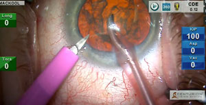
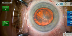
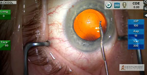
An eye with a shallow anterior chamber is at increased risk for endothelial cell loss during phacoemulsification. A convex anterior capsule increases the risk of
An eye with a shallow anterior chamber is at increased risk for endothelial cell loss during phacoemulsification. A convex anterior capsule increases the risk of
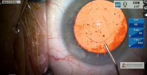
Determination of the correct IOL power for a highly myopic eye with posterior staphyloma is fraught with difficulty. Here I employ two methods that can
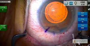
This patient has persistent negative dysphotopsia after cataract-implant surgery performed 18 months ago. A combination of techniques is used to achieve the desired reposition of

In this case I present a variety of phaco techniques and pearls as cataract extraction and multifocal IOL implantation is performed in an eye with
A very interesting case, indeed! Here we will see a patient with considerable head movement, very advanced corneal endothelial dystrophy, pseudoexfoliation, and zonular laxity that

This month’s case features methods for dealing with the problem of a patient with a very active Bell’s Phenomenon. The use and design of my
Following a standard cataract procedure, I address the reduction of IOP in this patient with open-angle glaucoma by implanting a stent in the trabecular meshwork.
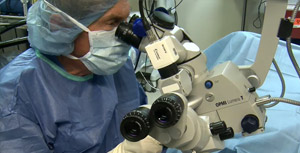
Many patients experience severe photophobia in response to the microscope light. These patients are also often hyperesthetic, and this patient presented with both issues. Here
This patient has a dense cataract, low ocular rigidity and unreliable preoperative biometry. The use of a high flow rate of 55 cc/min* to remove
In this issue I discuss and demonstrate the synergism of high IOP and high vacuum settings during phacoemulsification.
Tom is a courageous and engaging young man with advanced Duchenne’s Muscular Dystrophy. Years of steroid treatment have caused him to develop extremely dense cataracts
This patient underwent cataract-implant surgery 3 weeks ago, has severe negative dysphotopsia and, fortuitously, a mild hyperopic refractive error.However, the preexisting capsulorhexis is too small